venerdì 28 novembre 2008

Vasculitis / Behcet disease


Findings

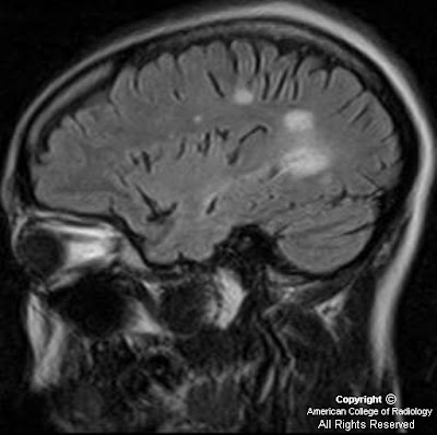
Figure 1, Figure 2, and Figure 3 demonstrate increased signal in the region of the bilateral basal ganglia, left thalamus, subthalamic region and brainstem. Diffusion-weighted images shows an area of restricted diffusion (Figure 2 and Figure).
Figure 4 shows contrast enhancement on T1WI in the left basal ganglia and focal meningeal enhancement.
Figure 5 single voxel MR spectroscopy within the region of interest demonstrates increased choline, slightly decreased NAA, and presence of lactate peak.
Figure 6, Figure 7, and Figure 8 Sagittal and axial T2 and axial T1 post contrast images show an abnormal long segment of high T2 signal in central cervical cord with subtle contrast enhancement.


Diagnosis: Vasculitis/Behcet disease


CNS vasculitis is a heterogeneous group of disorders characterized by nonatheromatous inflammation and necrosis of blood vessel walls. Arteries and veins are affected; it can involve intracranial vessels of any size.

A variety of systemic inflammatory diseases can cause vascular inflammation and stroke. Neuro-Behcet is a type of CNS vasculitis. Behcet disease is a chronic, relapsing, inflammatory disease characterized by presence of recurrent and usually painful mucocutaneous ulcers, genital lesions, ocular lesions, neurologic manifestations and cutaneous manifestations. Behcet is uncommon in the United States.

Neurologic manifestations are seen in approximately 10-25% of patients with Bechet disease. CNS manifestation results from arterial or venous thrombosis. Neuro-Behcet disease (NBD) usually shows three clinical patterns: A brainstem syndrome, a meningomyelitic syndrome and an organic confusion syndrome.

The typical MRI findings are multiple focal T2 signal abnormalities, restricted diffusion in the acute phase of disease, and patchy vascular and leptomeningeal enhancement. The most common site of involvement is the brainstem, followed by white matter, internal capsule and basal ganglia or thalamus. Brainstem atrophy is one of the manifestations of chronic NBD. Meningeal involvement is a less frequent finding. Follow up studies show change in site, size and shape of the lesions. Cerebral venous thrombosis is seen in approximately 0.6 to 10% of Behcet disease. The spinal cord involvement is rare. It usually shows long segment lesions demonstrating high signal in T2 WI, which may show patchy enhancement. Thoracic and cervical cords are usual sites of involvement.

In the largest series to date, the clinical features and outcomes of 200 patients with Behcet disease and neurologic involvement were reported. On average, a period of approximately 5 to 6 years elapsed between the onset of the earliest non-neurologic symptoms of Behcet disease and the appearance of neurologic symptoms or findings. In a small percentage of Behcet, neurologic findings may appear concurrently or precede non-neurologic features.

The prognosis varies with the type of neurologic process. Those with dural venous thrombosis or other non-parenchymal processes are less likely to have recurrent disease, disability, or premature death. By comparison, patients with parenchymal disease have a worse outcome.

giovedì 27 novembre 2008

CPPD of the cervical spine - "crown dens syndrome"


Findings

Figure 1: Sagittal view from a CT of the cervical spine utilizing a bone algorithm demonstrates a well-defined subchondral cyst in the odontoid process associated with partially calcified retro-odontoid soft tissue. There is also an erosion of the C5 spinous process as well as a fluid collection interposed between the spinous processes of C4 and C5. Calcification of the C2/C3 intervertebral disc is also noted. There is also minimal calcification of the longus colli.

Differential for crystal arthropathies that involve the cervical spine include
- CPPD
- Hydroxyapatite deposition disease
- Gout
- Other (rare)


Diiagnosis: CPPD of the cervical spine - "crown dens syndrome"


CPPD (calcium pyrophosphate deposition disease) is one of the entities known to cause crystal deposition in cartilage. It is most commonly found to be idiopathic, but other etiologies include a hereditary autosomal dominant condition, hyperparathyroidism, and hemochromatosis. While the classic locations for CPPD include the triangular fibrocartilage of the wrist, menisci of the knee, and pubis symphysis, the spine can also be involved. CPPD crystal deposition can be found in several structures of the spine including the ligaments, intervertebral discs, joint capsules, and synovium.

CT is the best modality to see the various calcific deposits in the cervical spine, while MRI is usually used to evaluate for spinal cord compression or myelopathy. Imaging findings include calcification of the intervertebral disc as well as disc space narrowing, “vacuum phenomena,” and vertebral sclerosis. Calcification in the periodontoid tissue in association with acute neck pain has been coined the “crown dens syndrome.” In addition to this acute symptomatology, these calcifications, particularly in the retro-odontoid region, can eventually lead to neurologic compromise from mass effect in the form of ventral cervicomedullary compression in the elderly. Deposition within the joints, such as the atlantoaxial and facet joints, can lead to erosions, subchondral cysts and pathologic fractures. CPPD deposits in the spinal ligaments can cause either a focal or diffuse enlargement of the ligaments and cause spinal stenosis.

The differential for calcific deposits in the cervical spine is a limited one. Hydroxyapatite deposition disease (HAAD) can present with periodontoid calcifications and can be indistinguishable from CPPD on CT. Patient history and, ultimately histological analysis of the crystals, help differentiate between the two. HAAD can also present as a calcific tendonitis with calcifications in the longus colli muscles, usually at the C1-C2 level. The inflammatory response to the CPPD deposits in the intervertebral disc space can be aggressive enough to mimic a discitis. Correlation with the patient’s clinical history would be helpful. In difficult cases, a biopsy may be needed. Gout can cause similar findings as CPPD including erosions of the cervical osseous elements, including the odontoid process, as well as mimic a discitis. Gout’s main imaging findings include erosions and proliferative osseous changes. Clinical history and/or crystal analysis may be needed for definitive diagnosis.

mercoledì 26 novembre 2008

Meningioma


Findings

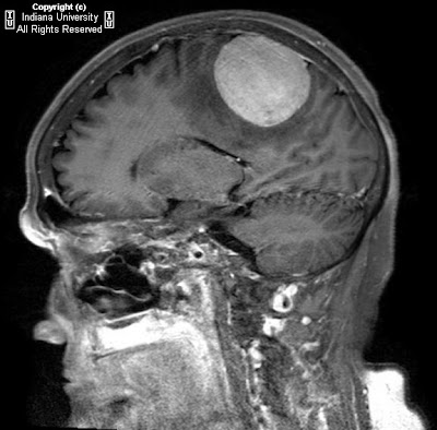
CT shows a large circumscribed vertex mass in the right parietal region with heterogeneous hyperdensity and calcification. There is significant vasogenic edema and minimal if any mass effect or midline shift.
MRI from next day shows broad attachment of the lesion to the dura with moderate contrast enhancement. T2 prolongation compatible with vasogenic edema is again present.

Differential diagnosis:
- Meningioma
- Metastatic disease with hemorrhage and/or calcification
- GBM
- Low grade astrocytoma
- Angiosarcoma
- Tuberculoma


Diagnosis: Meningioma


Discussion

Meningiomas are thought to arise from arachnoid cap cells and may arise in the spinal cord or intracranially. Fewer than 10% are symptomatic. They may present with headache, seizure, or focal neurologic signs due to cranial nerve or brain parenchymal compression or vascular compression. Known causes include radiation and genetic abnormalities (including a relationship to NF2). Other causes are speculated as well. Meningiomas are generally considered benign tumors. However, a few histologic types can break this rule and invade cortex and even metastasize. Therapy includes conventional surgery and radiosurgery. Chemotherapy can be used following resection. Angiography is often performed for surgical planning and occasional embolization.


Radiologic overview of the diagnosis

Plain films of the skull may demonstrate hyperostosis and increased vascular markings. CT and MRI demonstrate extra axial, dural based lesions. Meningiomas typically enhance homogeneously and may have an enhancing dural tail (which may be more evident on coronal or sagittal MRI depending on the location)..On CT, the lesion may be isoattenuating to hyper attenuating but may contain calcifications. Vasogenic edema will likely be present and may be more apparent on MRI. T1 and T2 signal is variable. MR spectroscopy demonstrates a high alanine peak. Buckling of the cortex (seen in this case) is strongly suggestive of an extra axial lesion and should narrow the differential diagnosis. Other clues to extra axial location are brain cysts and trapped CSF. Angiographic findings include a sunburst vascular pattern and "mother-in-law" blush (comes early and stays late).


lunedì 24 novembre 2008

Intracranial chondrosarcoma


Findings

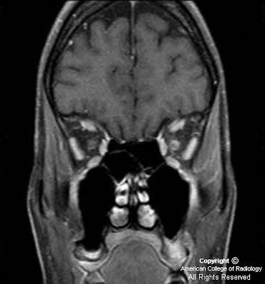
Figure 1: Sagittal T1 demonstrates a low-signal mass centered in the sphenoid bone and extending into the anterior cranial fossa. The pituitary and suprasellar cistern are preserved.
Figure 2: Coronal T2 image shows displacement of the frontal lobes without brain edema confirming extra-axial location. The mass is high signal on T2 images which is non-specific but typical of chondrosarcoma. There is edema in and around both optic nerves.
Figure 3 and Figure 4: T1 weighted fat-suppressed post gadolinium axial and coronal images demonstrate avid enhancement of the solid mass.
Figure 5 and Figure 6: Axial and coronal CT demonstrate the calcified matrix within the mass taking characteristic “ring and arc” shapes. The anterior and superior walls of the sphenoid sinus are eroded confirming the aggressiveness of the tumor.

Differential Diagnosis:
- Chondrosarcoma
- Meningioma
- Chordoma
- Metastasis
- Lymphoma


Diagnosis: Intracranial chondrosarcoma


Intracranial chondrosarcoma is a slow-growing, locally invasive, rare malignant neoplasm of cartilaginous origin which accounts for 0.15% of all intracranial tumors and most commonly affects the skull base. Often when encountered, other anterior skull base malignancies, such as meningioma, metastasis, chordoma, rhabdomyosarcoma, and lymphoma may be difficult to distinguish. However, recognizing the range of appearances of intracranial chondrosarcomas on various imaging modalities along with clinical, gross, and histological studies may allow for improvement in the diagnosis, evaluation, and management of this malignancy.

Primary chondrosarcoma is divided into multiple variants, depending on the location and histological characteristics. These include conventional, clear cell, myxoid, mesenchymal, extraskeletal, and dedifferentiated. Skull base chondrosarcomas are most commonly of the conventional type and occupy areas that include petrosal bone, temporoccipital bone, clivus, sphenoethmoidal complex, and less commonly the frontal, parietal, and ethmoidal bones. Conventional chondrosarcomas can be further classified into histological subtypes of grade I, grade II, and grade III, with grade I demonstrating the least malignant and aggressive potential. The mesenchymal variant is the most malignant of all types and typically presents in a younger population. This type has a predilection for dural and cerebral extension.

Intracranial chondrosarcomas have been shown to be minimally sensitive to conventional radiation therapy, thus requiring radical excision of the tumor for effective management. Therefore, diagnostic imaging with MRI and CT is significant for neurosurgical assessment of tumor invasion and its anatomic orientation with any surrounding vascular, bony, and neural structures. Radiological imaging has enhanced diagnosis as CT is useful in finely delineating tumor invasion, bony invasion, and abnormal “ring and arc” or stippled calcification typical of chondrosarcomas. The tumor is normally isoattenuated or hyperattenuated with some degree of heterogeneous enhancement. MRI with gadolinium allows for evaluation of significant vessels and nerves such as the carotid arteries and optic nerves that lie in the preferred area of tumor growth. Furthermore, the tumor appears with decreased signal on T1 weighted images and increased signal on T2-weighted images. MRI enhancement will be mild to moderate, typically described with a “honey-combing” appearance due to islands of cartilage. In addition, it has been demonstrated that there is only mild edema surrounding chondrosarcomas in contrast to other skull base malignancies. MRI perfusion has also been documented to be effective in differentiating chondrosarcomas from other anterior skull base malignancies by assessing the cerebral blood volume and perfusion. Tumor vascularity is variable depending on the histological type; however, conventional and mesenchymal chondrosarcomas are commonly hypovascular, which differentiates it from the significantly vascular meningioma and metastatic lesion.

Although rare, intracranial chondrosarcomas should be considered in the differential diagnosis of any skull base malignancy that causes cranial nerve deficits. Clinically, patients commonly present with headaches, tinnitus, dizziness, decreased sense of hearing, visual symptoms such as diplopia and other oculomotor disorders depending on the location of the mass. The mean age of patients with intracranial chondrosarcomas has been reported to be 37 without any gender predilection. Although conventional radiation has limited indication, there has been considerable debate regarding optimal treatment with proton radiotherapy in combination with surgical excision. The difficulty in evaluating the outcomes of these various treatment strategies for skull base chondrosarcomas stems from the fact that this is a rare malignancy.

Sturge-Weber syndrome


Findings

Gyriform calcifications are observed over the left occipital lobe in the CT exam. The MRI demonstrates atrophy of the left cerebral hemisphere. There is enlargement of the left choroid plexus which demonstrates homogeneous enhancement post contrast. There is also gyriform enhancement post contrast most significantly on the left occipital lobe. There is diffuse enhancement of the subcutaneous tissues over the left eye.


Diagnosis: Sturge-Weber syndrome


Key points

Classically the patients have a facial port-wine stain, ipsilateral intracranial abnormalities, contralateral hemiparesis, hemiatrophy, mental retardation, and homonymous hemianopia. The severity of these features varies widely patient to patient. Commonly the patients will have glaucoma on the affected side. Seizures are also very common.
Only 8% of patients with port-wine stains have Sturge-Weber Syndrome. 13% of Sturge-Weber syndrome patients do not have a facial angioma.
There is no clear genetic link at this time. There is no sex or race predilection and it is very seldom seen more than once in the same family. Several different chromosomal abnormalities have been implicated.
Radiographically one can see "tram track calcifications" which are leptomeningeal calcifications like those seen on the CT image.
MRI can demonstrate the angiomatous abnormalities. In this case the cutaneous capillary angioma (port-wine stain) is well demonstrated as is the meningeal angiomatosis over the left occipital lobe. Cerebral hemiatrophy is well demonstrated by MRI as is the choroidal angiomatosis.
Multiple therapies are employed in these patients. The port-wine stains can be "removed" with laser treatments. Seizures can be treated with anticonvulsants. Refractory seizures can be treated surgically. The surgeries can be as extensive as a hemispherectomy.


venerdì 21 novembre 2008

Acute necrotizing encephalitis (ANE)


Findings

Figure 2: Axial T2-weighted image showing bilaterally symmetric hyperintensity in the thalami. Note the target appearance of the lesions.
Figure 3: Axial T2-weighted image showing bilaterally symmetric hyperintensity in the dorsal pons.
Figure 4 and Figure 5: Coronal FLAIR images showing bilaterally symmetric hyperintensity in the thalami and dorsal columns.


Diagnosis: Acute necrotizing encephalitis


Acute Necrotizing Encephalitis (ANE) characteristically occurs in children after a mild antecedent illness. The peak age of incidence is six to eighteen months, however, ANE can occur in older children as well (the patient in this particular case was nine years old). Typically, the patient will present within a few days of a mild illness (e.g. fever and/or upper respiratory infection) with seizures, decreased level of consciousness, and/or vomiting.

Pathologic evaluation of the lesions demonstrates necrosis (due to severe edema) in the thalami, tegmentum, and dentate nuclei. In addition to necrosis, there is florid petechial hemorrhage around small parenchymal vessels. Pathologically, ANE is differentiated from ADEM by the lack of inflammatory cells found in ANE, as opposed to the presence of lymphocytes in ADEM. In addition to characteristic lesions of the thalami, tegmentum, and dentate nuclei; about one half of those affected will also have patchy lesions in the cerebral white matter (similar in appearance to ADEM). Unlike the thalamotegmental lesions, the patchy cerebral white matter lesions of ANE are not hemorrhagic. The presence of hemorrhage in patchy cerebral white matter lesions instead suggests the diagnosis of Acute Hemorrhagic Encephalomyelitis, which is distinct from ANE and ADEM.

Imaging studies in ANE typically show bilaterally symmetric lesions of the thalami, which may extend to the lateral putamina and external capsule, as well as tegmentum and cerebellar nuclei. The lesions are often necrotic and hemorrhagic. Ring-enhancement around the areas of hemorrhage and necrosis can be seen within a few days of onset. Initially, on diffusion-weighted imaging (DWI), the lesions exhibit restricted diffusion due to cytotoxic edema (restricted diffusion results in increased signal intensity on DWI). As necrosis develops, the movement of water molecules is less restricted (and ultimately unrestricted, as cell membranes dissolve), resulting in decreased signal intensity in areas of necrosis on DWI. Magnetic susceptibility artifact, secondary to hemosiderin deposition, may also contribute to the hypointensity seen in the center of the lesions on DWI; however, depending on the type of DWI employed, magnetic susceptibility artifact is probably less of a factor than decreased restriction of diffusion due to necrosis.

mercoledì 19 novembre 2008

Terson syndrome


Findings

Figure 1 and Figure 2: Axial noncontrast head CT demonstrates hyperdensity within the basal cisterns and sylvian fissures indicating diffuse subarachnoid hemorrhage.
Figure 3: Axial noncontrast CT with attention to the orbits demonstrates lentiform hyperdensity layering along the posterior aspect of the globes.


Diagnosis: Terson syndrome


Terson syndrome is defined by the presence of retinal, vitreous or any intraocular hemorrhage in association with subarachnoid hemorrhage. Several theories have been proposed with regard to its development. The most common theory suggests that raised intracranial pressures associated with subarachnoid hemorrhage leads to ocular venous outflow obstruction, thereby resulting in ocular hemorrhage. The hemorrhage is usually bilateral and composed of hyperdense crescentic collections along the posterior globe, near the optic nerve head. Other entities such as melanoma, metastatic lesions, and hemangiomas are in the differential, however in the setting of subarachnoid hemorrhage, Terson syndrome should be strongly considered.

Once findings of intraocular hemorrhage are noted, ophthalmologic consultation should be requested in order that a detailed fundoscopic exam be performed. Establishing a baseline is important to follow the progression or regression of the hemorrhage. Most hemorrhages clear spontaneously, however in those that do not, a vitrectomy may be needed to preserve vision.

giovedì 13 novembre 2008

Neurofibromatosis Type I (von Recklinghausen Disease)


Findings

FLAIR images (Figure 1, Figure 2, and Figure 3) show multiple hyperintense scattered foci in the cerebellum, medial temporal lobes, bilateral basal ganglia, corpus callosum and cortex/subcortical regions (Figure 1 and Figure 3). Figure 2 demonstrates an abnormally enlarged optic chiasm.
Postcontrast images (Figure 4, Figure 5, and Figure 6) show heterogeneous enhancement of these lesions. The right cerebellar lesion showed increase in size on follow up imaging and on biopsy proved to be low grade astrocytoma.
Figure 7: Bilateral optic nerves and optic chiasm are markedly enlarged, consistent with optic gliomas.


Diagnosis: Neurofibromatosis Type I (von Recklinghausen Disease)


Neurofibromatosis Type I (NF 1) is the most common neurocutaneous disorder, with an incidence of 1 per 3,000 births. NF1 is transmitted as an autosomal dominant disorder, with 50 % of cases representing spontaneous mutations. The gene locus for NF 1 is on Chromosome 17 with the gene product, neurofibromin, thought to function as tumor suppressor gene.

The CNS manifestations of NF 1 area
- Foci of abnormal signal intensity (FASI) – Present in 60-85 % of NF 1 patients. Non enhancing foci of T2 prolongation, thought to represent areas of myelin vacuolization. These are most commonly seen in the globus pallidi, cerebellum, brainstem, internal capsules, centrum semiovale and corpus callosum. These foci wax for 2-10 years and usually wane by 20 years of age. FLAIR is the best sequence for detection and enhancement in these lesions is worrisome.
- Optic nerve gliomas (ONG) - Seen in 15-20%. Usually low grade and more commonly bilateral. Optic nerve/chiasm involvement is more common than posterior extension. ONGs are associated with higher incidence of brain parenchymal tumors.
- Brain tumors- Astrocytomas are seen in 1-3% of NF 1 patients. Common sites are brainstem/cerebellum and splenium of corpus callosum. These tumors have an earlier presentation, are more likely multicentric and are much less aggressive than their sporadic counterparts. MR Spectroscopy may show increased choline in both FASI and tumors, but NAA is near normal in FASI while tumors show decreased levels. Persistent enhancement and increasing size are worrisome features for tumors. There is slight increased incidence of medulloblastoma and ependymoma in NF 1 patients as well.
- Plexiform neurofibromas (PNF) - Common locations are orbit, scalp and skull base. Enlarged optic foramina and fissures (ONG) and foramen ovale (PNF) may be seen. Paraspinal PNFs are also common. Degeneration to neurofibrosarcoma is seen in 2-12% of cases.
- Sphenoid wing dysplasia
- Spine- Dural ectasia, lateral thoracic meningocele, scoliosis
- Ocular findings- Lisch nodules (Iris hamartomas), Buphthalmos, pthisis bulbi.

Extra spinal ependymoma at level of coccyx, subcutaneous type


Findings

A well-defined low density mass which is adherent to the coccyx bone and extends posteriorly to the skin. No rectal invasion. No bony destruction.

Differential diagnosis
- Sacrococcygeal teratoma.
- Lipoma.
- Pilonidal cyst.
- Extraspinal ependymoma
- Coccygeal chordoma
- Rhabdomyosarcoma


Diagnosis: Extra spinal ependymoma at level of coccyx, subcutaneous type


Discussion

Ependymomas are the most common tumors of glial origin in the spinal cord and are usually located in the cauda equina or filum terminale. Extra spinal medullary ependymomas are rare. There are two types, the subcutaneous type and the presacral type. The subcutaneous type can occur due to residual ependymal tissue present at the coccygeal medullary vestige, which is a remnant of the caudal portion of the neural tube. This ependymal-lined structure is present at the post-anal pit, in the subcutaneous soft tissues over the end of the coccyx. This patient has a subcutaneous type. The presacral type is thought to arise from extradural remnants of the filum terminale or as an extension of intradural filum terminale.

This patient had the myxopapillary type of tumor, which is the most common subcutaneous type of extra spinal ependymoma. Papillary and subependymal types also exist. Immunohistochemical stains can aid in the diagnosis with positive stains for glial fibrillary acidic protein, S-100 and vimentin.

These tumors are slow-growing and tend to present late, with a mean age of 17 years of age. There is equal male and female distribution. The mass is usually asymptomatic, located at the intergluteal fold. These masses are frequently misdiagnosed as a pilonidal cyst.

Surgical resection is curative. The survival rate is 85-100%. Metastases occur in about 20%, including pulmonary, pleural, osseous, and inguinal nodal metastases have been documented. Distant metastases can occur up to 10-20 years after the initial presentation. For presacral ependymomas, local recurrence occurs in 71% if the coccyx is not excised. Radiotherapy is recommended for incomplete resection or local metastases. Chemotherapy is not currently recommended.

mercoledì 12 novembre 2008

Acute left MCA stroke with thrombus seen on CT-angiogram


Findings

Non-contrast head CT shows a left MCA stroke with a hyper dense linear structure in the left MCA. Angiogram confirms suspicion of thrombus in the left MCA.


Diagnosis: Acute left MCA stroke with thrombus seen on CT-angiogram


Key points

On MRI: Diffusion restriction with correlation on ADC map is diagnostic.
Seen as a hypo attenuating lesion on non-contrast Head CT.
Head CT can have false negative for acute stroke.
The hyperdense MCA sign implies acute thrombus and is a poor prognostic indicator.

venerdì 7 novembre 2008

Multiple sclerosis (MS)


Findings

34 yo) Figure 1: Sagittal FLAIR image delineates septo-callosal interface hyperintensities, perpendicular periventricular hyperintensities extending into the deep white matter, and a juxtacortical lesion.
Figure 2: Coronal fat saturated T1 post gadolinium demonstrates enhancement of the left optic nerve.

38 yo)Figure 3: Sagittal FLAIR (3a) and axial FLAIR (3b) images demonstrate confluent periventricular and juxtacortical oval plaque-like hyperintense lesions perpendicular to the ventricular axis known as “Dawson’s fingers”.

44 yo) Figure 4: Axial FLAIR shows hyperintense plaques with one extending to a juxtacortical location. Figure 5: Sagittal T2 shows hyperintense plaques within the brainstem and upper cervical spinal cord. Figure 6: Sagittal FLAIR demonstrates periventricular and subcortical hyperintensities.


Diagnosis: Multiple sclerosis


Multiple Sclerosis (MS) is a demyelinating inflammatory CNS disorder of unclear etiology. It specifically affects oligodendrocytes, thus eliminating their supportive function to the neurons they serve. Women are affected twice as frequently as men, usually between the ages of 20 and 40 years. MS is a clinical diagnosis based on history, neurological examination, and paraclinical studies including MR imaging, evoked potentials and CSF studies. The hallmark of this disease is its dissemination in space and time. Diagnosis of MS separates it from clinically isolated syndromes (CIS). MS has different subtypes, the most common being the relapsing-remitting type. Other subtypes include primary progressive, progressive relapsing, and malignant/Marburg. Related demyelinating processes such as Schilder’s diffuse sclerosis and Balo’s concentric sclerosis may be considered MS subtypes. However, recurrent optic neuritis and neuromyelitis optica (Devic’s disease) have been shown to be distinctly separate entities.

Specific MR imaging characteristics include the presence of T2 hyperintensity at the septo-callosal interface and ovoid lesions perpendicular to the ventricles, known as Dawson fingers. These occur along the deep medullary veins. Active lesions may enhance avidly or poorly depending on the degree of acuity and severity. Lesions may also involve the cortex, juxtacortical white matter, brainstem, and spinal cord. These lesions, given their protean distribution and overall presentation, have an extensive differential diagnosis and therefore multiple criteria schemes have been developed to aid in the diagnosis of MS. The differential diagnosis of MS includes acute disseminated encephalomyelitis and its possible subtypes of neuromyelitis optica (Devic disease), acute optic neuritis, and acute transverse myelitis; microvascular white matter ischemic changes; progressive multifocal leukodystrophy; neurosarcoidosis; hypertensive encephalopathy; vasculitis; and encephalitis.

The 2001 International Panel on the Diagnosis of Multiple Sclerosis (IPDMS) (McDonald et al.) “McDonald Criteria” require objective evidence of CNS lesions disseminated in space and time in order to diagnose MS. Spatial criterion is defined by the Barkhof-Tintore MR imaging criteria, which require three of the following four findings: 1) at least one gadolinium-enhancing lesion or 9 T-2 hyperintense lesions; 2) at least one infratentorial lesion; 3) at least one juxtacortical lesion; 4) at least 3 periventricular lesions. Lesions should be greater than 3-mm in cross-section. A spinal cord lesion may substitute for a brain lesion, and in the setting of oligoclonal IgG bands or elevated IgG/Albumin ratio in the CSF, only 2 instead of 9 T2 lesions are needed to satisfy the criteria. Temporal criterion is satisfied by follow up imaging 3 or more months after the onset of the clinical event. The 2005 IPDMS (Polman et al.) suggested revisions to the 2001 McDonald criteria based on several research studies which followed. These modifications were made to allow for the following: 1) multiple spinal lesions may be used to substitute for brain and infratentorial lesion criteria, provided that they are more than 3mm in size, the length is less than 2 vertebral body heights, and the lesion occupies only a portion of the cord cross section, 2) an enhancing spinal cord lesion may be substituted for an enhancing brain lesion, and 3) for dissemination in time, a new T2 lesion discovery interval may be reduced from 3 months to 1 month. Polman also suggested that CSF studies are no longer needed in order to consider a diagnosis of primary progressive MS.


The 2001 IPDMS (McDonald et al.) “McDonald Criteria”

Dissemination in Space: 3 of the following 4
- At least 1 gadolinium-enhancing lesion or 9 T2-weighted hyperintense lesions*
- At least 1 infratentorial lesion
- At least 1 juxtacortical lesion
- At least 3 periventricular lesions

* + oligoclonal IgG bands or elevated IgG/Albumin ratio in the CSF, only 2 T2 lesions are needed


Dissemination in Time: One of the following
- A new enhancing lesion at least 3 months after the initial clinical event in a new clinically relevant area
- A new T2 lesion identified on a new MRI study at least 3 months after the initial scan


The 2005 IPDMS modified “McDonald Criteria”, (Polman et al.)

Dissemination in Space: 3 of the following 4
- At least 1 gadolinium-enhancing lesion (spinal cord, brainstem, and brain) or 9 T2-weighted hyperintense lesions (spinal cord, brainstem, and brain)*
- At least 1 infratentorial lesion (spinal cord, brainstem, and cerebellum)
- At least 1 juxtacortical lesion
- At least 3 periventricular lesions

* +oligoclonal IgG bands or elevated IgG/Albumin ratio in the CSF, only 2 T2 lesions are needed


Dissemination in Time: One of the following
- A new enhancing lesion at least 3 months after the initial clinical event in a new clinically relevant area
- A new T2 lesion identified on a new MRI study at least 1 month (30 days) after the initial scan


Conclusion

Diagnostic criteria for MS and its variants are continually revised as more data becomes available and MR imaging technology improves. Notwithstanding, attention to detail with precise temporospatial descriptions of lesions is essential for the diagnosis of MS and may also have prognostic value.