mercoledì 30 aprile 2008

Extrapontine myelinolysis (osmotic demyelination syndrome)


Findings

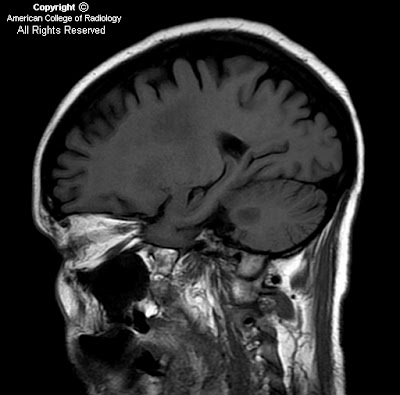
Figure 1: Sagittal T1 image taken parasagitally at the level of the dentate nucleus. There are T1 hypointensities within the subcortical white matter and in the brachium pontis.
Figure 2: Axial T2 at the level of the cerebral subcortical white matter. There is T2-hyperintensity in the centrum semiovale bilaterally. The signal abnormality is fairly symmetrical.
Figure 3: Axial T2 at the level of the splenium of the corpus callosum. There are T2-hyperintensities within the splenium of the corpus callosum and asymmetrical T2-hyperintensities along the corticospinal tracks within the posterior limbs of internal capsules.
Figure 4: Axial T2 at the level of the middle cerebellar peduncles. There is T2-hyperintensity within the brachium pontis bilaterally. Note that the signal abnormality is symmetrical. Also note that the signal within the pons was normal.
Figure 5: Axial FLAIR at the level of the splenium of the corpus callosum. There are FLAIR-hyperintensities within the splenium of the corpus callosum and asymmetrical FLAIR -hyperintensities along the corticospinal tracks within the posterior limbs of internal capsules.
Figure 6 and Figure 7: Axial diffusion weighted and ADC map images at the level of the middle cerebellar peduncles. There is increased signal in both DWI and in the ADC map, consistent with the subacute stage of myelinolysis (four weeks after onset of symptoms). The DWI and ADC maps at the level of the centrum semiovale and splenium (not shown) demonstrated similar findings.


Diagnosis: Extrapontine myelinolysis (osmotic demyelination syndrome)


Central pontine myelinolysis (CPM), was first described in 1959 in patients with a history of alcoholism and malnutrition. Extrapontine myelinolysis (EPM) is the extrapontine manifestation of osmotic myelinolysis and can occur with or without central pontine myelinolysis. In one autopsy series, myelinolysis was found to be confined to the pons (CPM) in about 50% of cases, combined CPM and EPM occurred in about 30 % of cases, and exclusive EPM occurred in about 20% of cases7.

CPM and EPM are characterized by symmetrical loss of myelin with relative preservation of axons and neuronal cell bodies. Pathologically, there is dissolution of the myelin sheaths of fibers. There is however, no evidence of inflammation. The myelinolysis occurs with relative sparing of the nerve cells and axon cylinders.

Although initial reports were largely confined to chronic alcoholics, osmotic myelinolysis has also been seen in patients with electrolyte disturbances, particularly hyponatremia that has been rapidly corrected (more then 10mmol/l/day), and in liver transplant patients being immune-suppressed with cyclosporine. Alcohol continues to be a particularly frequent etiology (up to 40% of cases)7.

Osmotic myelinolysis can affect a variety of sites resulting in a variety of symptoms. The MRI appearance is characteristic with lesions being symmetrical. However, the timing of the appearance of lesions on MRI may be significantly delayed. If there is a strong clinical suspicion of the diagnosis, repeat imaging at 1 to 2 weeks may reveal lesions that were not initially apparent. Diffusion weighted imaging (DWI) might have the capability of detecting lesions undetectable on conventional MRI earlier, with one case report in the literature showing an altered DWI in a patient within 24 hours of symptoms at a time when conventional MRI findings were inconspicuous.

The prognosis of the osmotic demyelination syndrome is variable. In a reported series, about 6 % of patients died, about 32 % survived but were left dependent, about 32% had some deficits but were independent, and about 30% recovered completely. Initial clinical features are not predictive of outcome and a recent case series found conventional MRI findings were not prognostic but the prognostic role of diffusion-weighted imaging is still unclear 3,4,8.

In conclusion, CPM and EPM are the same disease, sharing the same pathology, associations, and time course but differing in clinical manifestations due to differences in the affected structures. Both diseases have lesions that are strikingly symmetrical.

martedì 22 aprile 2008

CNS arteriovenous malformation (AVM)


Findings

There is a large, diffuse left cerebral hemisphere arteriovenous malformation, which is located mostly within the posterior left frontal lobe. The remainder of the brain parenchyma appears within normal limits. On the MR angiogram, there is a large left frontal and parietal lobe mass of tangled vessels supplied mostly by left MCA, but also left ACA branches. On the conventional angiogram, there is a tangle of vessels in the left parietal lobe, with arterial supply from the prefrontal and post frontal branches of the Rolandic division. There is venous drainage drainage into the superior sagittal sinus and sphenoparietal sinus.

Diagnosis: CNS arteriovenous malformation


Key points

Classic imaging finding is "bag of worms" from flow voids.
Minimal or no mass effect.
Strong enhancement on CT.
MRI characteristics varies with flow rate and presence / age of hemorrhage.
Peak presentation between 20 and 40 years old.
50% will hemorrhage; 25% will have seizures.
Rare spontaneous regression.
Treatment includes embolization, radiosurgery, and also surgical resection.


mercoledì 16 aprile 2008

Choroid plexus carcinoma


Findings


Figure 1: Axial T2 WI shows a large, heterogeneous, lobulated, frond-like lesion within the left lateral ventricle with a prominent flow void suggestive of a vessel within the lesion. There is diffuse dilatation of the ventricular system.
Figure 2: Axial T2 weighted image demonstrates another heterogeneous lesion in the right Foramen of Luschka.
Figure 3 and Figure 4 : Axial and coronal T1 fat suppressed post gadolinium MR images demonstrate heterogeneous enhancement within the mass lesions with diffuse leptomeningeal enhancement in the interpeduncular and perimesencephalic cisterns. Parenchymal invasion is also seen with enhancing foci in the left occipital region.
Figure 5: Axial T1 fat suppressed post gadolinium MR image through the abdomen demonstrates circumferential leptomeningeal enhancement around the cord.


Diagnosis: Choroid plexus carcinoma


The choroid plexus is the neuroepithelial tissue responsible for the production of CSF within the cerebral ventricular system. Neoplasms of the choroid plexus are uncommon tumors which can form anywhere there is choroid in the ventricular system. These tumors occur in proportion to the amount of normally present choroid tissue. The lateral ventricle is the most common site (50% of cases), followed by the fourth ventricle (40%) and the third ventricle (5%). About 5% of choroid plexus tumors arise in more than one location.

Choroid plexus tumors usually present in the first decade of life. Most choroid plexus tumors (about 80%) occur as the benign, slowly growing papilloma, a WHO grade I tumor with a favorable overall prognosis. The malignant variant manifests as a much more biologically aggressive WHO grade III tumor, and is far more common in children than adults.

Clinical presentation is usually due to increased intracranial pressure and hydrocephalus, due mainly to CSF overproduction, but mechanical obstruction and impaired CSF resorption may also contribute.

Choroid plexus papillomas usually present as lobulated intraventricular masses with intense, homogeneous enhancement in a child. These tumors are soft, well-circumscribed cauliflower-like masses with prominent lobulations peripherally. The mass can be hyperattenuating on CT with calcification seen in 25%. Necrosis resulting in heterogeneity and parenchymal invasion are characteristic features for choroid plexus carcinoma. Extension from one ventricle to another or into the cerebellopontine angle is a characteristic feature. The presence of focal neurological signs also suggests choroid plexus carcinoma due to parenchymal invasion. CSF seeding can be seen with both papilloma and carcinomas and enhanced MRI of the entire neuraxis is recommended prior to surgery to evaluate extent of disease.

Choroid plexus cyst


Findings

The patient has an approximately 6-7 mm choroid plexus cyst.


Diagnosis: Choroid plexus cyst


Discussion

Choroid plexus cysts are a relatively frequent finding on obstetrical sonographic imaging with an incidence of 0.2 to 2.5%. They are thought to occur due to folding of the neuroepithelium with trapping of secretory products and desquamated cells. On histological evaluation of these choroids plexus cysts, there is no epithelial lining, and they are usually filled with clear colored fluid which may have some debris.

The importance of recognizing this finding is related to its association with karyotypic abnormalities, most commonly trisomy 18. Almost 40-70% of patients with trisomy 18 (Edward syndrome = constellation of findings including micrognathia or facial cleft, omphalocele, clenched hands with overlapping index fingers, rocker-bottom feet, small cerebellum with prominent cisterna magna) have choroid plexus cysts which often times are bilateral and larger than 1 cm in diameter. However, in the absence of any other anatomical abnormalities, less than 1% of fetuses with choroid plexus cysts will have trisomy 18. There is also a reported association of choroid plexus cysts with trisomy 21 (up to 17%) although less commonly than with trisomy 18.

Management of the pregnancy upon detection of these cysts is controversial. Almost 90% of choroids plexus cysts resolve by the 28th week of gestation. In over 95% of the patients, there is no clinical significance. However, the decision to perform amniocentesis is based on the results of the serum triple screen results (Edward syndrome = low maternal AFP, low HCG, low estiol), maternal age, and the presence of any other anatomic abnormalities. In the presence of any other anatomic abnormality and choroid plexus cysts, there is a 4% incidence of Edward syndrome. Some obstetricians will order follow-up obstetrical ultrasounds to evaluate for resolution of these cysts. However, even if the choroid plexus cysts resolve, there is no effect on the reduction of likelihood of karyotypic abnormalities.


Radiologic overview

Ultrasound is the widely accepted standard radiological imaging modality for obstetrical fetal anatomy survey. Part of the standard set of sonographic images obtained during the fetal intracranial survey include an "axial" image plane through the lateral ventricles for purposes of ventricular measurement to exclude hydrocephalus. The choroid plexus is often the most echogenic structure within the head. Choroid plexus cysts most often appear as well circumscribed anechoic structures within the brightly echogenic choroid plexus tissue located in the atria of the lateral ventricles. Identification of these cysts should prompt a thorough sonographic evaluation for other potential anomalies including an enlarged cisterna magna or myelomeningocele, omphalocele, or extremity malformations.

Choroid plexus cysts can also be seen on fetal MRI examinations. On MR, these cysts will follow fluid signal intensity which will be low on T1 and bright on T2 image sequences.

giovedì 10 aprile 2008

Osteoblastoma


Findings

Figure 1: Axial CT image of the cervical spine with bone windows demonstrates a round, well-defined sclerotic lesion in the right C5 facet.
Figure 2 and Figure 3: Coronal and sagittal MIP CT images demonstrate that the expansile portion of the lesion involves the transverse process, facet and lamina with sparing of the vertebral body.

Differential diagnosis of posterior element lesions
- Aneurysmal bone cyst
- Hemangioma
- Osteoid osteoma/osteoblastoma
- Giant cell tumor


Diagnosis: Osteoblastoma (path proven)


Osteoblastomas are benign osteoid forming lesions which commonly occur in the spine (42%). They are differentiated from osteoid osteomas by their larger size. Osteoblastoma presents as an expansile, geographic lesion with a sclerotic margin occurring in the posterior elements. The lesions may be centered in the pedicle, lamina, transverse or spinous process. Osteoblastomas may exhibit a range of densities varying from a lucent, mixed or blastic appearance. This accounts for the range of appearances on T1 and T2 weighted MR images. Because they are osteoid-forming lesions, osteoblastomas demonstrate increased uptake on bone scan.

Although osteoblastomas usually present with a narrow zone of transition with surrounding sclerosis, they may present with aggressive features with a wide zone of transition and cortical breakthrough. Prostaglandins released by the lesion may cause extensive peritumoral edema. This peripheral inflammatory response may result in periosteal reaction in the adjacent ribs, pleural thickening or effusion and ossification of the ligamentum flavum.

Patients with osteoblastomas usually present in their 2nd to 3rd decade of life (90%). Osteoblastoma is a differential diagnostic consideration of “painful scoliosis.” The most common signs and symptoms include dull pain with scoliosis concave to the side of the lesion. Neurologic symptoms may arise from compression of the cord or nerve roots. Treatment of the lesion involves curettage with bone graft. Occasionally, radiofrequency ablation is used. However, the proximity of the lesion to the cord often limits the use of this treatment. Preoperative embolization is sometimes employed as well.

Central neurocytoma


Findings

There is an enhancing heterogeneous suprasellar mass adjacent to the third ventricle. Mass effect on the cerebral peduncles and lateral and third ventricles.

Differential Diagnosis:
- Neurocytoma
- Subependymoma
- Craniopharyngioma
- Low-grade glioma


Diagnosis: Central neurocytoma


Key points

Typically supratentorial, intraventricular, bubbly, well circumscribed mass with low grade enhancement. Calcification is present 50-70%. (this patient does not demonstrate some of these features).
Extraventricular extension is a poor prognostic sign (as in this case).
Often attached to septum pellucidum.
Commonly cause obstructive hydrocephalus due to obstruction of foramen of Monro.
CT typically shows mixed solid and cystic architecture with rare hemorrhage.
Decreased metabolism on PET.
<1% of intracranial neoplasms.
Present from age 20 to 40 with signs of increased intracranial pressure.
Subependymoma may be radiographically indistinguishable, but is typically in those older than 40 and is more commonly related to the 4th ventricle (whereas CN involves the 4th ventricle extremely rarely).

martedì 8 aprile 2008

Paraneoplastic limbic encephalitis (PLE)


Additional clinical history: 50-year-old female with a history of small cell lung cancer (SCLC) presents with a history of worsening short term memory over the last 3 months.


Findings

Figure 1: Axial FLAIR MR image demonstrates increased signal in the left medial temporal lobe, uncus and hippocampus.


Diagnosis: Paraneoplastic limbic encephalitis (PLE)


Paraneoplastic limbic encephalitis (PLE) is a remote neurologic effect of cancer outside of the CNS. It is the most common of the clinical paraneoplastic syndromes. The typical presenting symptoms include short-term memory loss, seizure, or psychologic features such as mood or behavioral disturbance.

The most frequent neoplasms associated with PLE are SCLC, testicular tumors, breast cancer, Hodgkin’s lymphoma, and thymoma. SCLC makes up the majority of cases. Up to 60% of patients do not have the diagnosis of cancer at presentation, with neurologic symptoms preceding the discovery of tumor by weeks to months.

The etiology involves immune mediated injury by autoantibodies or cytotoxic T-cells. Pathological studies reveal neuronal loss, perivascular inflammatory infiltrates and microglial nodules in affected limbic structures, without cancer cells.

Serologic markers for PLE are anti-Hu in small cell lung cancer and anti-Ta in patients with testicular cancer. Identification of these markers coupled with the characteristic MRI finding helps establish the diagnosis.

Typical MRI findings include unilateral or bilateral mesial temporal lobe hyperintensity on T2 weighted images or FLAIR. On T1 sequences, the mesial temporal lobes may be hypointense. Herpes simplex encephalitis may have similar findings in early stages of disease. However, the patients usually develop mass effect and signs of edema. Herpes encephalitis is an acute febrile illness whereas PLE is a subacute illness. This fact makes clinical correlation particularly important.

Treatment is primarily at targeting the primary malignancy.

Takayasu’s arteritis


Findings

The MRA demonstrates focal stenoses of the right innominate artery, proximal right subclavian artery, and proximal left common carotid artery with post-stenotic dilatation. There is also marked narrowing of the right internal carotid artery and focal stenosis of the proximal left subclavian artery before the left vertebral, with reconstitution distal to the left vertebral. The CT angiogram again demonstrates segmental large vessel vascular stenoses and left common carotid fusiform aneurysm.

Differential Diagnosis:
- Takayasu's arteritis
- Temporal arteritis
- Atherosclerosis
- Syphilitic aortitis
- Fibromuscular dysplasia
- Idiopathic arteritis


Diagnosis: Takayasu’s arteritis


Key points

Defined as a granulomatous inflammation affecting predominately elastic arteries (aorta, main aorta branches, and pulmonary arteries). This is the only aortitis that causes stenosis or occlusion of the aorta.
Most commonly involves left subclavian, left common carotid, brachiocephalic trunk, renal arteries, celiac trunk, SMA, and pulmonary arteries.
Affects females more often than males (8:1). More often affects oriental persons aged 12-66.
For the first few months to years, the patient experiences a prepulseless phase that has fevers, sweats, myalgias, weight loss, and arthralgias, The pulseless phase then shows signs of limb ischemia and renovascular hypertension. Patients often have an ESR > 20 mm/hour.
Acutely there is granulomatous infiltration of the elastic fibers of the media that progresses to a fibrotic stage over weeks to months. The fibrosis leads to constriction from proliferation, thrombosis, and aneurysm formation.
Treat with steroids, and then angioplasty after active inflammation subsides.
Angiography (and CT angiography) may demonstrate wall thickening, stenoses, occlusions, luminal and mural calcium and thrombus deposition.

Classification
- Type I: is the classic pulseless type involving the subclavian, carotid, and brachiocephic trunk
- Type II: is a combination of types I and III
- Type III: is the atypical coarctation type involving the descending thoracic and abdominal aorta with major branches
- Type IV: is the dilated type with dilatation of the aortic length and its branches

mercoledì 2 aprile 2008

Temporal bone fracture


Findings

There is air in the infratemporal fossa(Figure 1). There is opacification within the right sphenoid sinus (Figure 2, Figure 3, and Figure 4). There is a longitudinal temporal bone fracture (Figure 2, Figure 3, and Figure 4).


Diagnosis: Temporal bone fracture


Blunt force trauma to the skull base is the most common cause of temporal bone fracture. The location of the injury determines the orientation of the fracture line in relation to the petrous ridge:
- fractures coursing parallel to the petrous ridge are classified as longitudinal
- fractures coursing perpendicular to the petrous ridge are classified as transverse

Longitudinal fractures account for 80% of all temporal bone fractures. Longitudinal fractures are typically sustained as a result of blunt trauma to the temporoparietal region. The fracture line runs along a lateromedial axis. It originates in the squamous temporal bone and runs along the external auditory canal, coursing towards the middle ear space. The otic capsule is typically spared in a longitudinal fracture. However, conductive hearing loss may occur as a result of ossicular discontinuity, hemotympanum, or tympanic membrane perforation. Sound will lateralize to the affected ear on the Weber test.

Transverse fractures account for 20% of all temporal bone fractures. They occur as a result of blunt trauma to the occipital skull. The fracture line runs along a posteroanterior axis. It originates in the region of the foramen magnum and courses anteriorly, through the otic capsule. Disruption of the otic capsule results in sensorineural hearing loss. On the Weber test, sound will localize to the normal ear. Transverse fractures are associated with a greater incidence of complications, including facial nerve paralysis, nystagmus, and cerebrospinal fluid leak.

Temporal bone fractures are often identified on routine computed tomography scans of the head in the setting of trauma. However, high-resolution CT (HRCT) with thin-cut sections in the axial and coronal planes best evaluates the extent of injury to the temporal bone.


Longitudinal

Frequency: 80%
Orientation to petrous ridge: Parallel
Axis of fracture line Posteroanterior: Mediolateral
Hearing loss: Conductive
CN VII complications: 6-13%


Transverse

Frequency: 20%
Orientation to petrous ridge: Perpendicular
Axis of fracture line Posteroanterior: Posteroanterior
Hearing loss: Sensorineural
CN VII complications: 30-50%

Glioblastoma multiforme ("Butterfly glioma")


Findings

CT shows a large ill-defined area of hypodensity and mass effect in the right frontal lobe which extends across midline. MRI confirms a large heterogeneously enhancing bilobed mass crossing the rostrum of the corpus callosum. There is associated vasogenic edema and sulcal effacement. Mass effect is seen on the lateral ventricles. Mild midline shift. There is restricted diffusion within the right frontal lobe mass.

Differential Diagnosis:
- Glioblastoma multiforme (GBM)
- Primary CNS lymphoma
- Metastasis
- Demyelinating disease
- Infection


Diagnosis: Glioblastoma multiforme ("Butterfly glioma" distribution)


Key points

Most common primary malignant intracranial neoplasm (50-60% of astrocytomas).
Most commonly in cerebral hemispheres, but can be seen in brainstem and cerebellum (which is more common in children).
May invade meninges and cause dural enhancement.
Often demonstrates a marked mass effect, marked surrounding edema and a necrotic core.
Demonstrates irregular rim enhancement on CECT and MR + C.
"Butterfly Glioma" (such as this case) involves both hemispheres and the corpus callosum.
May be sporadic or associated with a tumor syndrome including NF1, p53 mutation, Turcot syndrome, Ollier Disease, and Maffucci syndrome.
20% are multifocal.